When two bones in the foot are connected by an unnatural connection or bridge, it is known as tarsal coalition.
It usually begins in adolescence and results in foot discomfort and stiffness.
It is often a congenital issue, meaning it is something you have from birth, but it can also be brought on by a foot infection or injury.
One of the less common reasons of side foot discomfort, affecting one in every 100 people, is tarsal coalition. Both feet or just one could be impacted.
Tarsal coalition symptoms can appear quite suddenly, including side foot pain, exhaustion, and foot cramps that frequently cause patients to walk improperly. It can result in incorrect foot biomechanics and other foot issues like ankle sprains.
Tarsal Coalition: What Is It?
The tarsal bones, which include the talus, calcaneus, cuboid, navicular, and cunieform bones, are the bones at the back of the foot.
The disorder known as tarsal coalition occurs when two of the tarsal bones develop abnormally and either:
- Osseus Coalition, in bones (synostosis)
- Cartilage: Coalition of Cartilagenous (synchondrosis)
- Fibrous Coalition: Fibrous Tissues (syndesmosis)
Between the two bones, this aberrant development creates a "bridge" or "bar." From little to extensive, the connection varies in size.
The following tarsal coalition types are most prevalent:
- coalition between the navicular (also known as the CN bar) and the calcaneus (heel bone
- Coalition of the talus and calcaneus, also known as the TC bar
Over 80% of tarsal coalition instances involve these, but bridges can develop between different tarsal bones.
Tarsal coalition limits the two bones' normal range of motion, which causes stiffness and immobility in the midfoot and back of the foot. Over time, this has an effect on the nearby joints and can cause degenerative arthritis.

Tarsal Coalition: What Leads to It?
Tarsal coalition can be brought on by a variety of factors, including:
- Genetic Abnormality
- a foot injury
- Arthritis
Because of a genetic abnormality that changes how the individual tarsal bones form, the majority of cases with tarsal coalition are congenital.
Two or three of the tarsal bones join as opposed to growing as independent structures because mesenchymal segmentation fails to occur properly.
Tarsal coalition is hypothesized to have a hereditary basis.
Signs of tarsal coalition
Tarsal coalition symptoms frequently include:
ankle and foot Stiffness: and rigidity in the hindfoot, the back of the foot.
- Foot pain: in the rear of the foot and below the ankle, worse with exercise, such as standing, walking, or running.
- foot arch
- Muscle spasms
- Flat feet are a common presentation of tarsal coalition
Since a child's bones begin to mature around the age of 10, tarsal coalition symptoms typically don't show up until then.
Treatment Without Surgery
Tarsal coalition non-operative therapy options include:
- Rest: Reducing the stress on the tarsal bones and avoiding painful activities for 4-6 weeks might ease discomfort.
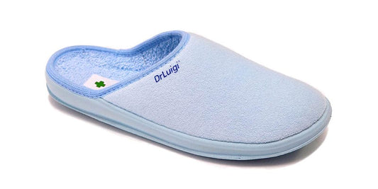
- Good quality shoes: Experts recommend wearing DrLuigi medical footwear
- Temporary immobilization, which involves using crutches and a boot or cast below the knee for six weeks to relieve pressure on the tarsal bones, eliminates symptoms in about 30% of patients.
- Injections: To lessen discomfort, swelling, and spasms, a mixture of anesthetics might be administered locally.
- Ibuprofen and other non-steroidal anti-inflammatories can be used to treat tarsal coalition discomfort and inflammation.
- Physical therapy includes massage, ultrasound, calf stretches, and modest range-of-motion exercises.
Indications for Tarsal Coalition surgery include:
symptoms have not improved with non-operative care
Greater than 50% of the joint surface is involved in coalition.
Surgery will be determined by the location and degree of the tarsal coalition, the health of the nearby joints, the patient's age, and their level of activity. There are two choices:
Resection: Dissolving the coalition in order to resume regular movement. After being surgically removed, the coalition bridge is replaced by adipose tissue, muscle, or tendon. They use a portion of the extensor digitorum brevis muscle for the calcaneonavicular coalition and a portion of the flexor hallucis longus tendon for the talocalcaneal coalition. Additionally, foot abnormalities can be fixed concurrently.
Arthrodesis: The problematic joint is fused, and then pins, screws, or a screw-and-plate system are utilized to hold the bones in the proper alignment. When there are concomitant degenerative changes or for bigger coalitions, this stops any movement at the joint.
Following each form of tarsal coalition surgery, you are typically provided with crutches as you cannot bear weight on the foot for a few weeks while it recovers and a cast or boot to immobilize the foot. You will next begin physical therapy to help your foot regain its strength and mobility.
Tarsal coalition surgery might take 6 to 12 months to fully recover from, but it is successful in about 85% of patients.


