The foot is the lower extremities most extreme component. It contains incredibly complicated anatomical components and serves as the body’s sole support when standing or walking. The foot’s muscular structures are divided into two groups: those that belong to the foot and those that belong to the lower leg.
Bones of the feet
The foot skeleton is separated into three sections. The bones of the toes (ossa tarsi), the bones of the middle part of the foot (ossa metatarsi), and the bones of the root of the foot (ossa tarsi) (ossa digitorum pedis). Phalanges are the components that make up the bones of the toes.
The tarsus, or root of the foot, is made up of seven bones arranged in two rows, proximal and distal. The talus (ankle bone), heel bone (calcaneus), and navicular axis make up the proximal, or upper, group (conical bone). Three cuneiform bones (ossa cuneiforma) and the cuboid bone make up the distal or lower group of bones. All the bones are arranged in a single row.
The only bone directly connected to the lower leg bones is the talus, or ankle bone. It is divided into three sections. These are the head of the bone (caput tali), the neck (collum tali) and the trunk (corpus tali) (corpus tali). The ankle bone is formed in the 8th week of pregnancy and is especially important for strengthening the ankle joint. Fracture of the ankle neck occurs during excessive plantar flexion. In this case, it is possible to move the body of the talus backwards.
The calcaneus is the largest bone in the root of the foot. It has a square shape. It is the only bone of the foot that touches the ground when walking and standing. The back of the heel bone has an enlarged tubercle (tuber calcanei) (tuber calcanei).
Ossa metatarsalia are the bones of the middle foot. There are 5 metatarsal bones.
The bones of the toes (ossa digitorum pedis) are made of phalanges.
Sesame bones (ossa sesamoidea) are also interesting in the anatomical structure of the feet. Sesame bones do not occur in all people and are clinically relatively insignificant. The most famous sesame bone is the patella in the knee.

Ankles
The lower leg and foot are connected by the lower and upper ankle. The upper ankle joint is called the articulatio talocruralis, and the lower talocalcaneonavicular. The metatarsophalangeal joint (articulationes metatarsophalangeal) is the joint between the joints of the toes and the bones of the central part of the foot. In addition to these three joints in the foot, there are many other joints.
The upper ankle joint (articulatio talocruralis) consists of the lower ends of the tibia and fibula. The other part of the joint is the trochlea tali. a hollow in the ankle bone (talus) containing the articular surface.
The lower ankle joint (articulatio talocalcaneonavicular) connects the talus (ankle bone) and the calcaneus (heel bone) (heel bone). The movements in the lower ankle joint are closely related to the movements in the central part of the foot, and the articular surfaces themselves allow slipping and rotation.
The ankle joint is the joint in the body most affected by injuries. Mid-body sprains (medial sprains) are rare as the joint is firmly fixed in this area by the deltoid ligament
Metatarsophalangeal joint is clinically very important due to numerous clinical conditions such as hallux valgus, gout, osteoarthritis… The first metatarsophalangeal joint is particularly affected. In hallux valgus, it deforms and enlarges, and this is associated with the displacement of the big toe towards the lateral. The condition is more common in women, especially older women, and in people who wear uncomfortable and inappropriate shoes. Such shoes have a pointed toe and a high heel. Fortunately, this condition can be prevented with wide and comfortable footwear, which is increasingly following fashion trends on the market, and is therefore more acceptable to people of lower ages, where prevention makes the most sense.

The muscles of the foot are divided into two large groups, such as the muscles of the back of the foot and the muscles of the sole.
There are two small muscles on the back of the foot, which are: m. extensor digitorum brevis te m. extensor hallucis brevis. The first muscle works by extending the 2nd to 4th toes, and the second by extending the big toe. Both muscles are innervated n. ischiadicusom tj. its branch n. fibularis profundus.
The sole muscles are divided into three parts: medial, lateral, and central.
Connective envelopes of the feet
The connective tissue of the foot plantation is called the fascia plantaris. Contains depth and surface sheet. Deep is called lamina profunda, and superficial aponeurosis plantaris.
The aponeurosis plantaris is a solid connective sheath and is separated from the skin by adipose tissue. Participates in the maintenance of the arches of the feet. The parts of the plantar aponeurosis are central, medial, and lateral.
Plantar fasciitis is treated with stretching exercises and night splints, and in case of failed surgery.
Blood vessels of the feet
The most important artery of the foot is a. dorsalis pedis. It is a direct branch of a large blood vessel. tibialis anterior which supplies the tibia. The dorsalis pedis artery lies directly on the back of the foot and is covered with fascia and skin. Follow the nerve n. fibularis profundus. The mentioned artery can also be palpated, and this is done when checking the pulsations on the foot. People with impaired circulation will have a weakened pulse or will not even be able to palpate it. These are often patients with diabetic foot or chronic wounds. Further a. dorsalis pedis divides into aa. tarsal mediales, a. tarsalis lateralis, a. arcuate, a. plantaris profundus the aa. metatarsals dorsales.
Furthermore, the a. Plantaris medialis (medial plantar artery) and the a. Plantaris lateralis are the terminal branches of the posterior tibial artery and participate in the supply of the plantar.
The more prominent veins of the feet are v. Dorsalis pedis, vv. tibiales anteriores te vv. tibiales posters.
Lymph is described along with blood vessels, but foot lymph is relatively insignificant.
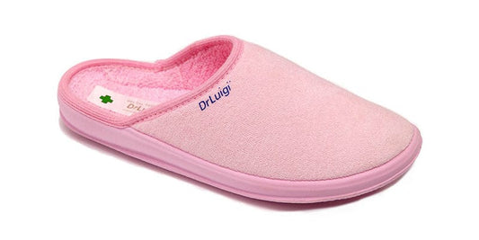
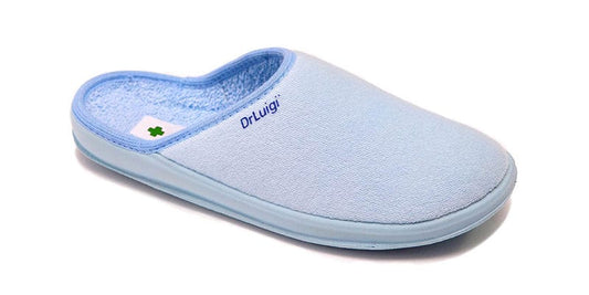
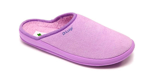
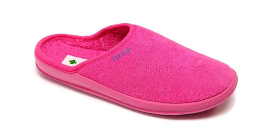
It is especially important to emphasise the importance of orthopaedic footwear for people with impaired circulation. Patients with impaired circulation must be careful when choosing footwear, as they are at increased risk of developing trophic ulcers. Footwear must be breathable, made of natural materials and comfortable. Any pressure from shoes can make wounds harder to heal than in healthy people. DrLuigi footwear follows these high standards and is especially adapted for patients with diabetes, dysfunctional circulation… It is light, adaptable to each patient and highly functional.
Foot nerves
The N. ischiadicus is the largest nerve in the human body, extending from the pelvis all the way to the tips of the toes. It is as thick as a thumb, and branches to n. tibialis te n. fibularis communis. Sometimes these two nerves are already separated in the pelvic area so I n. ischiadicus does not even exist.


